

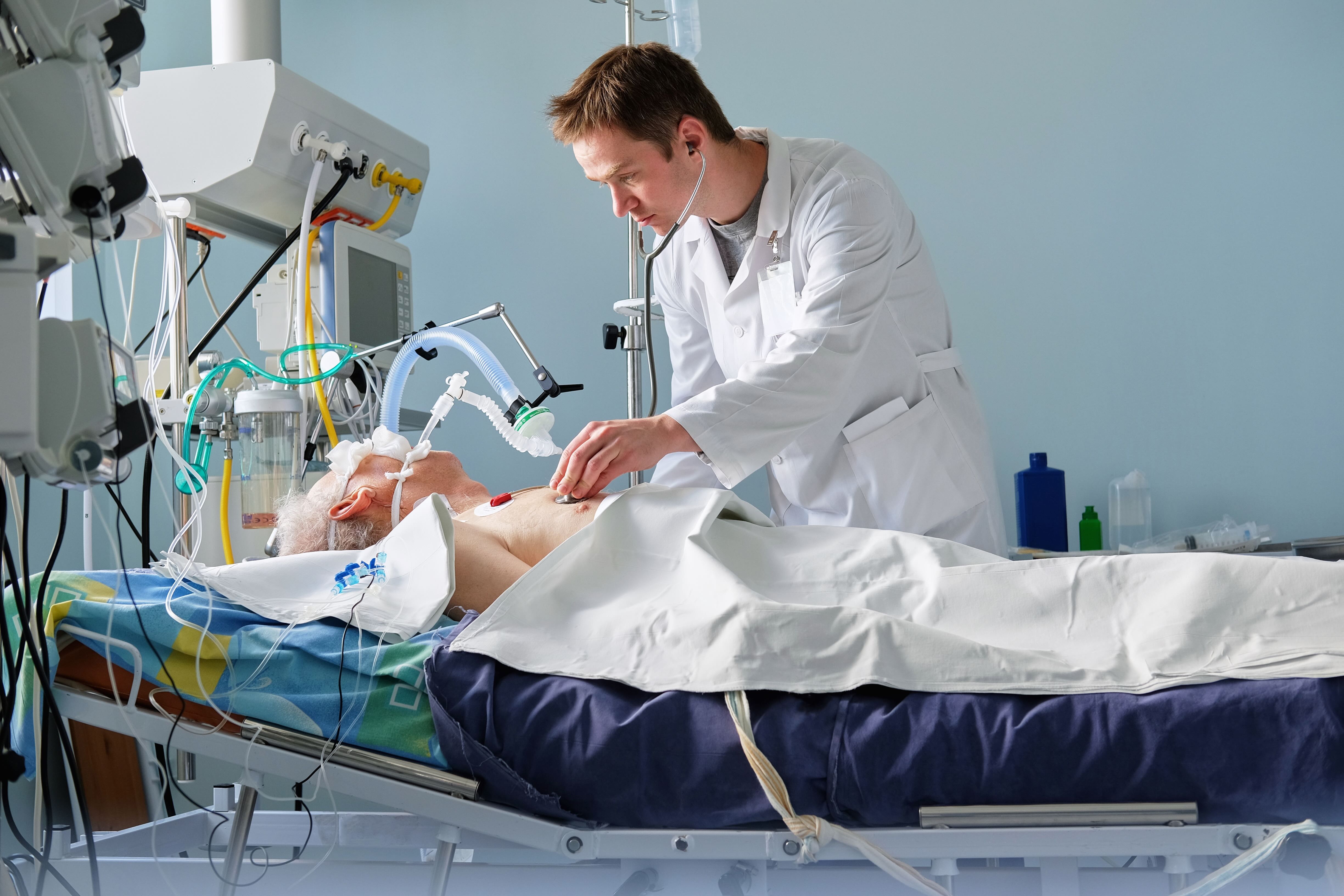
Mechanical ventilation saves lives, but it can also cause harm. Each patient's lungs respond differently to the same ventilator settings - what helps one patient may injure another. Yet clinicians must make these critical decisions with limited visibility into what's actually happening inside the lung. The result: preventable damage, prolonged ICU stays, and outcomes that depend too much on individual expertise rather than objective data.
Aerogram creates a patient-specific digital twin from routine imaging, then simulates how that individual lung responds to different ventilator settings. Clinicians receive objective guidance that reflect individual mechanical behavior for the first time - no guesswork, no trial and error.

Clinician orders an Aerogram for the patient.

AI generates a personalized digital twin if the patient's lung, extracting the 3D lung structure and pathology from the CT scan to build a functional replica.

Aerogram computes personalized ventilation maps by systematic testing of ventilator setting combinations on the digital twin.

Clinicians use the maps to navigate to personalized protective settings.

Enabling an improved patient trajectory.
Aerogram combines physics-based lung modeling, computational physiology, and AI to generate high-fidelity Digital Twins of each patient's lung from a routine CT scan.


Based on years of physics-based modeling research at the Technical University of Munich, including dozens of peer-reviewed, international, disciplinary as well as interdisciplinary journal publications, Aerogram computes clinically validated, personalized, actionable insights essential to improve patient outcomes.
Studies have validated the technologicy, including regional predictions of lung mechanics and inhaled drug transport & deposition against established imaging modalities like EIT and 3D SPECT.
